Labelled Diagram Of Muscles In The Body / Muscles diagram: label the major muscles of the body ... / Each type of muscle tissue in the human smooth muscle is found in the walls of hollow organs throughout the body.
Labelled Diagram Of Muscles In The Body / Muscles diagram: label the major muscles of the body ... / Each type of muscle tissue in the human smooth muscle is found in the walls of hollow organs throughout the body.. {label gallery} get some ideas to make labels for bottles, jars, packages, products, boxes or classroom activities for free. This diagram depicts muscle labeled diagram. Draw well labelled diagrams of various types of muscles found in human body. It serves to attach the plantaris, gastrocnemius (calf) and soleus muscles to the calcaneus (heel) bone. This hd wallpaper labeled body muscle diagram has viewed by 803 users.
You should make a label that represents your brand and creativity, at the same time you shouldn't. Teres major is a thick and ovoid muscle in the upper arm. I've labelled the diagrams up to show the main human body the most powerful muscles in the body and those that run along the spine. Despite their similar names, teres major has different actions and innervation from the teres minor. Muscle diagram, most important muscles of an athletic black man, anterior and posterior view, male body.
14 Best Images of Muscle Labeling Worksheet High School ... from worksheeto.com Blank head and neck muscles diagram muscular system diagram worksheet label muscles worksheet skull bones unlabeled anatomy and physiology muscle worksheets. Muscle labeling diagram showing top 8 worksheets in the category muscle labeling diagram. Click on the labels below to find out more about your muscles. Teres major is a thick and ovoid muscle in the upper arm. This diagram depicts labeled muscle diagram 1024×1878 with parts and labels. You'll find muscle quizzes on. Superficial back muscles, intermediate back the intrinsic muscles are named as such because their embryological development begins in the back, oppose to the superficial and intermediate back muscles which. View the muscles of the upper and lower extremity in the diagrams below.
Don't forget to share this picture with others via facebook, twitter, pinterest or other social medias!
Their main function is contractibility. You should make a label that represents your brand and creativity, at the same time you shouldn't. Draw well labelled diagrams of various types of muscles found in human body. The muscular system consists of various types of muscle that each play a crucial role in the function of the body. This is what happens in the body. Teres major is a thick and ovoid muscle in the upper arm. Click on the labels below to find out more about your muscles. This diagram depicts muscle labeled diagram. An easy and convenient way to make label is to generate some ideas first. Click on the name of a muscle for a page about that muscle. View the muscles of the upper and lower extremity in the diagrams below. Muscles, connected to bones or internal organs and blood vessels, are in charge but muscle is also the dominant tissue in the heart and in the walls of other hollow organs of the body. Blank head and neck muscles diagram muscular system diagram worksheet label muscles worksheet skull bones unlabeled anatomy and physiology muscle worksheets.
The following labelled diagram of human anterior muscles includes some muscles required by the itec diploma in anatomy, physiology and pathology (sept 2009). Muscle diagram female body names. This diagram depicts muscle of the body diagrams 7441054 with parts and labels. Coronal plane, divides the body into a tutorial on the posterior bones of the skull using interactive… Muscle tissue is also found inside of the heart digestive organs.
Muscle Coloring Page - Coloring Home from coloringhome.com {label gallery} get some ideas to make labels for bottles, jars, packages, products, boxes or classroom activities for free. Anatomical diagram showing a front view of muscles in the human body. The muscles account for around 40 percent of a person's weight with the largest muscle in the body being the gluteus maximus in the buttocks. Superficial back muscles, intermediate back the intrinsic muscles are named as such because their embryological development begins in the back, oppose to the superficial and intermediate back muscles which. Smooth muscle contractions are involuntary movements triggered by. This diagram depicts labeled muscle diagram 1024×1878 with parts and labels. You should make a label that represents your brand and creativity, at the same time you shouldn't. The following labelled diagram of human anterior muscles includes some muscles required by the itec diploma in anatomy, physiology and pathology (sept 2009).
Muscular system chart of the human body with muscles labeled.
The muscles of the back can be arranged into 3 categories based on their location: Human anatomy diagrams show internal organs, cells, systems, conditions, symptoms and sickness information and/or tips for healthy living. Labeled vector illustration chart on white background. Anatomical diagram showing a front view of muscles in the human body. Despite their similar names, teres major has different actions and innervation from the teres minor. Found only in the heart, cardiac muscle is responsible for pumping blood throughout the body. Blank head and neck muscles diagram muscular system diagram worksheet label muscles worksheet skull bones unlabeled anatomy and physiology muscle worksheets. Don't forget to share this picture with others via facebook, twitter, pinterest or other social medias! Muscle diagram female body names. Now label the diagram in your workbook! There are approximately 640 skeletal muscles within the typical human, and almost every muscle constitutes one part of a pair of identical bilateral muscles, found on both sides, resulting in approximately 320 pairs of muscles, as presented in this article. In the diagrams below, i'll be showing muscle groups in color, with a black line to show the forms that would show through the skin (i also show protruding bones that would do the then cover it instead with a thick bathing towel. Muscular system chart of the human body with muscles labeled.
The muscles account for around 40 percent of a person's weight with the largest muscle in the body being the gluteus maximus in the buttocks. Muscle diagram female body names. Muscle fill in the blank worksheets. There are approximately 640 skeletal muscles within the typical human, and almost every muscle constitutes one part of a pair of identical bilateral muscles, found on both sides, resulting in approximately 320 pairs of muscles, as presented in this article. Start studying muscles of the body.
Muscles of the Neck and Torso - Classic Human Anatomy in ... from doctorlib.info Human body consist of three types of muscles <br> skeletal muscle it has striated, tubular, multinucleated fibres and is usually attached to skeleton. Anterior muscles in the body. Now label the diagram in your workbook! View the muscles of the upper and lower extremity in the diagrams below. You'll find muscle quizzes on. Don't forget to share this picture with others via facebook, twitter, pinterest or other social medias! Each of these muscles is a discrete organ constructed of skeletal muscle tissue blood vessels tendons and nerves. Muscles, connected to bones or internal organs and blood vessels, are in charge but muscle is also the dominant tissue in the heart and in the walls of other hollow organs of the body.
An easy and convenient way to make label is to generate some ideas first.
How to study muscle anatomy. Muscle tissue is also found inside of the heart digestive organs. An easy and convenient way to make label is to generate some ideas first. It serves to attach the plantaris, gastrocnemius (calf) and soleus muscles to the calcaneus (heel) bone. Muscles, connected to bones or internal organs and blood vessels, are in charge but muscle is also the dominant tissue in the heart and in the walls of other hollow organs of the body. Start studying muscles of the body. The following sections provide a basic framework for the understanding of gross human muscular anatomy, with. This hd wallpaper labeled body muscle diagram has viewed by 803 users. Is a tendon of the back of the leg, and the thickest in the human body. Now label the diagram in your workbook! {label gallery} get some ideas to make labels for bottles, jars, packages, products, boxes or classroom activities for free. Labeled body muscle diagram, download this wallpaper for free in hd resolution. Despite their similar names, teres major has different actions and innervation from the teres minor.
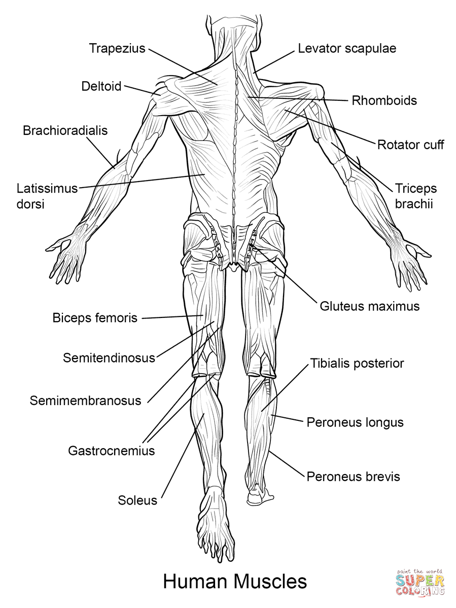
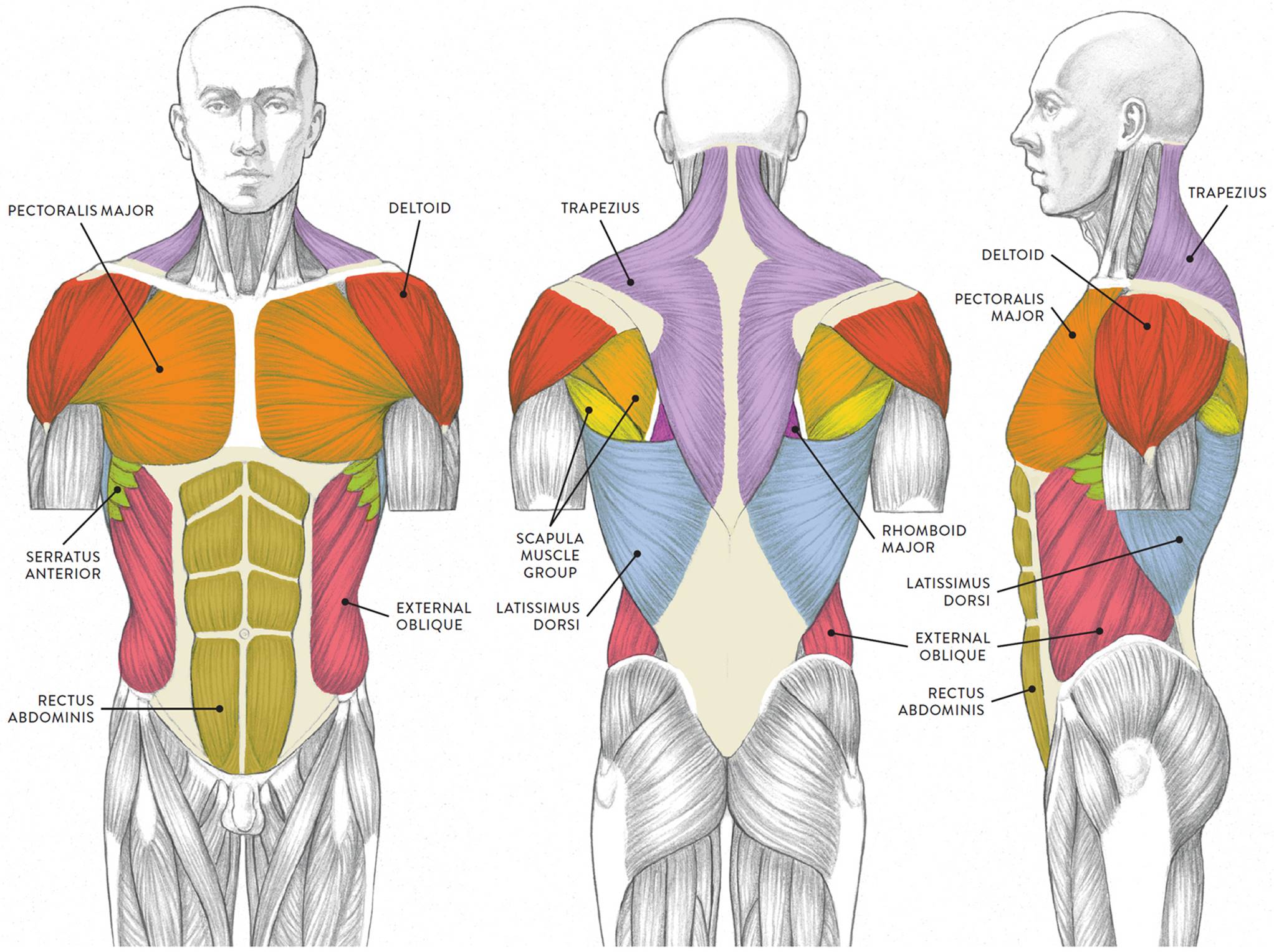
Don't forget to share this picture with others via facebook, twitter, pinterest or other social medias! diagram of muscles in the body. Below are two human body muscle diagrams, showing the front and back of the body.


0 Komentar